- Approximately 20 cm in length
- Starts at the cricoid cartilage and extends to the stomach
- Esophagus is composed of different types of muscles
- Proximal third contains striated muscle
- Distal third contains smooth muscle
- Middle section is composed of both striated and smooth muscle
- Esophageal motility - swallowing
- Initially there is pharyngeal contraction that transfers the "bolus" of solid or liquid material to the relaxed upper esophageal sphincter
- As material passes the sphincter muscles, the sphincter muscle contracts initiating a primary peristaltic wave that forces the material downward
- Secondary contractions occur along the lining of the esophagus in response to residual solid or liquid material that may still be in the esophagus
- Achalasia
- Loss of peristalsis within the muscles of the esophagus and failure of the lower esophageal sphincter to relax
- This results in prolonged retention of the food/liquid in the distal third of the esophagus
- Diffused esophageal spasm syndrome
- Results in spasms of the lower 2/3 of the esophagus and included intermittent chest pain or dysphasia
- This results in prolonged transit times and reduced esophageal emptying
- In addition, material within the esophagus moves within all three segments
- Nutcracker esophagus
- Exemplified by high amplitude peristaltic contractions in the body of the esophagus along with chest pain and/or dysphasia
- Food/liquid are retained within the mid to distal third of the esophagus
- Abnormal esophageal transit times have been associated with all connective tissue disorders that involve smooth muscle and striated muscles
- Smooth muscle disease Scleroderma, systemic lupus erythematosus, and Raynauds disease
- Striated muscles disease dermatopolymyositis
- Transit patterns show stagnation of food/liquid in the lower 2/3 of the esophagus
- Patient should be NPO after 12M
- Radiopharmaceutical
- Prepare 2 to 3 syringes with 15 mL water boluses in each
- Each bolus should contain 300 μCi of Tc99mSulfur Colloid
- Patient instruction (is very important)
- The patient will be given a syringe full of radiowater that will be placed in the patients mouth
- The patient will be asked to keep the radiowater in his/her oral cavity (dont swallow until I tell you)
- When acquisition is started, the patient will be asked to swallow the bolus and then continue to dry swallow for an additional 10 minutes
- Patient positioning
- In the supine position place the camera so that the detector is in an anterior position
- The field of view should include the oral cavity to the base of the stomach
- Acquisition parameters
- Two sets of acquisition should be completed for each bolus administered
- Matrix should be set at 64 by 64
- Collimator LEPH Gap or HS
- First set of acquisitions - 0.1 to 0.25 seconds per frame for 10 to 20 seconds
- Second set of images can be taken at 1 second frame for up to 10 minutes
- Following the swallow of the bolus, the patient should dry swallow every 10 to 15 seconds, until the study is done
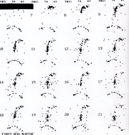
- The above image identifies the first set of images collected at 0.1 seconds per frame. Note the lack of activity seen in these images.
- Image processing
- ROIs may be drawn in several ways
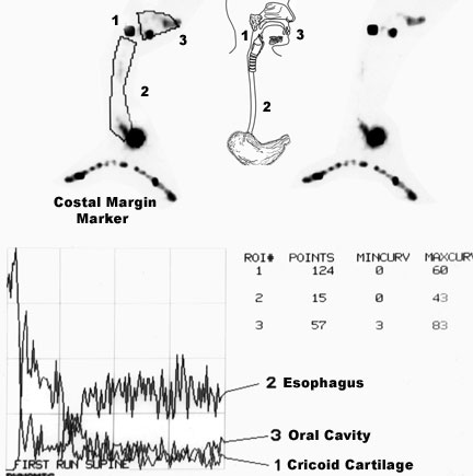
- Note that the images above indicate three ROIs mouth, cricoid cartilage, and the entire esophagus
- This data represents the initial images that were collected above
- Time activity curves are then generated
- According to the data above the bolus of activity arrives at the stomach within a second (normal) and no reflux or delayed transit is noted
- Modification of the ROIs
- Three ROIs can be drawn around esophagus to include upper, mid, and lower portions (1/3 each)
- This adds to the sensitivity of the exam by noting where the abnormal transit times might occur
- Consider the diseases described above and determine what might be the behavior of the radioactive bolus
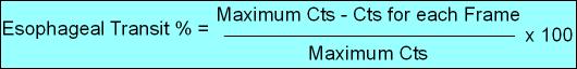
- Finally, a percent transit time can also be calculated using the above formula
- Maximum counts represent the frame where the initial bolus is at maximum counts in the esophagus (as its passing through)
- "Counts At End Frame" represent the activity remaining after a period time (t)
- Which frame do you select for t?
- Remember liquid should travel down the esophagus in no more than 1 second. After that time, any residual activity represents the inability of the esophagus to get the bolus to the stomach
- Hence, the frame you want to selecting is going to be after 1 second
- Therefore the "End Frame" represents the % traveled through the esophagus or %Esophageal Transit
- Note - The frame that you select after 1 second will represent residual activity remaining the esophagus
Return to the beginning of the document
Return to the Table of Contents